An FDG PET CT Scan is a diagnostic imaging test that helps doctors examine metabolic activity in different parts of the body. It combines Positron Emission Tomography (PET) and Computed Tomography (CT) to provide information about tissue activity and anatomical structures.
The scan uses a small amount of radioactive glucose-based tracer called 18F-Fluorodeoxyglucose (18F-FDG). It is commonly used in cancer evaluation, including staging, treatment response assessment and checking for suspected recurrence.
Patients looking for information about an FDG PET CT Scan in Navi Mumbai can learn about the procedure, preparation requirements, medical uses and safety considerations before undergoing the examination.
What Is an FDG PET CT Scan?
An FDG PET CT scan is a nuclear medicine imaging procedure that combines two imaging techniques to assess the body.
PET (Positron Emission Tomography)
Detects metabolic activity in tissues using the injected 18F-FDG radiotracer.
CT (Computed Tomography)
Provides detailed images of organs, bones and other anatomical structures.
The FDG tracer behaves similarly to glucose and accumulates in tissues according to their metabolic activity. Many cancer cells use glucose at a higher rate than surrounding tissues, making certain tumours visible on PET images.
However, increased FDG uptake is not specific to cancer. Infection, inflammation and normal tissue activity can also cause increased tracer uptake.
The combined PET and CT images help doctors locate areas of abnormal metabolic activity and interpret them alongside the patient's medical history and other investigations.
Why Is an FDG PET CT Scan Done?
An 18F-FDG PET CT Scan is primarily used in the evaluation and management of several types of cancer. The examination may be recommended for different clinical purposes.
1. Cancer Staging
FDG PET CT helps assess the extent of certain cancers by identifying metabolically active disease in the primary tumour, lymph nodes or other organs.
The results support treatment planning when PET CT is appropriate for the particular cancer type.
2. Monitoring Cancer Treatment Response
Doctors may recommend FDG PET CT during or after chemotherapy, immunotherapy or other cancer treatments to evaluate changes in tumour metabolic activity.
The timing of the scan depends on the cancer type and treatment received.
3. Detecting Suspected Cancer Recurrence
An FDG PET CT scan may help investigate suspected cancer recurrence when symptoms, laboratory findings or other imaging tests raise concern.
4. Evaluating Selected Tumours
In certain clinical situations, FDG PET CT helps assess metabolically active lesions and may assist in selecting an appropriate site for biopsy.
5. Other Medical Applications
FDG PET CT is also used in selected cases involving infection, inflammation, cardiac viability assessment and neurological disorders.
These applications require specific imaging protocols and clinical indications.
Which Cancers Can Be Evaluated Using FDG PET CT?
FDG PET CT is commonly used in the evaluation of several cancers, depending on their type, stage and clinical characteristics.
| Cancer type | Potential role of FDG PET CT |
|---|---|
| Lung Cancer | Staging, assessment of disease spread and suspected recurrence |
| Lymphoma | Initial staging and treatment response assessment |
| Head and Neck Cancer | Staging and evaluation of suspected residual or recurrent disease |
| Colorectal Cancer | Assessment of suspected recurrence or selected metastatic disease |
| Breast Cancer | Evaluation in selected advanced or recurrent cases |
| Oesophageal Cancer | Staging and treatment planning in appropriate patients |
| Melanoma | Assessment of advanced disease and suspected spread |
| Cervical Cancer | Staging and recurrence assessment in selected cases |
Important: FDG PET CT is not equally useful for every cancer. Some tumours show low FDG uptake, while others require different imaging techniques or specialised PET tracers.
The referring specialist determines whether FDG PET CT is appropriate for the patient's diagnosis.
How Is an FDG PET CT Scan Performed?
The FDG PET CT procedure generally involves the following steps.
Step 1: Patient Preparation and Blood Sugar Check
Before the examination, the imaging team reviews the patient's medical history, medications and preparation instructions.
Blood glucose is usually checked before administering the FDG tracer because elevated glucose levels can affect scan quality.
Step 2: FDG Tracer Injection
A small amount of 18F-FDG is administered through an intravenous injection, usually into a vein in the arm.
The tracer circulates through the bloodstream and is taken up by tissues according to their metabolic activity.
Step 3: Tracer Uptake Period
After the injection, the patient generally rests quietly for approximately 45–60 minutes.
Movement, talking and muscle activity are minimised during this period to reduce unnecessary tracer uptake in muscles.
Step 4: PET CT Imaging
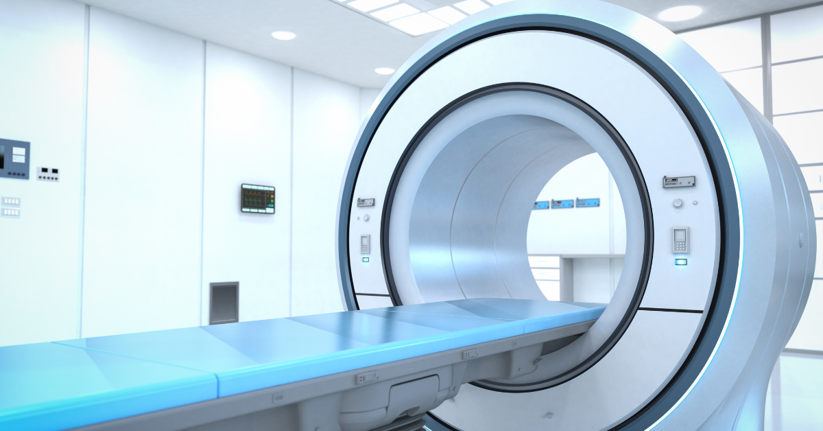
The patient lies on a scanning table that moves through the PET CT scanner.
The equipment acquires CT and PET images, which are combined to show anatomical structures and metabolic activity.
Patients are usually asked to remain still during image acquisition.
Step 5: Image Interpretation
A qualified nuclear medicine physician interprets the scan findings along with relevant clinical information and previous investigations.
The report is shared according to the imaging facility's reporting process.
How to Prepare for an FDG PET CT Scan
Following preparation instructions is important because food intake, physical activity and blood glucose levels can influence FDG distribution.
Before Your Scan
Do not stop prescribed medicines or change insulin doses without instructions from your treating doctor or imaging team.
How Long Does an FDG PET CT Scan Take?
An FDG PET CT appointment commonly takes approximately 2–3 hours, although the duration varies according to the imaging protocol and individual requirements.
This typically includes:
Additional images, delayed scanning or contrast-enhanced CT may increase the appointment duration.
Is an FDG PET CT Scan Safe?
FDG PET CT is an established diagnostic imaging procedure performed using controlled amounts of radioactive tracer and CT imaging.
The examination involves exposure to ionising radiation from both the PET radiotracer and the CT component.
The medical team considers the expected diagnostic benefit and radiation exposure before performing the scan.
Important Safety Considerations
Radiation exposure
The radiation dose varies depending on the tracer activity and CT scanning protocol.
Pregnancy
FDG PET CT is generally avoided during pregnancy unless the clinical benefit justifies the potential risk.
Breastfeeding
Special instructions may be required following tracer administration.
Diabetes
High blood glucose can affect FDG uptake and image interpretation. Individual preparation is important.
Contrast-related risks
If intravenous CT contrast is required, additional assessment may be necessary for allergies, kidney function and other relevant conditions.
An FDG PET CT scan should be performed when clinically indicated and interpreted by an appropriately qualified medical professional.
What Happens After an FDG PET CT Scan?
Most patients can resume normal activities after the examination unless they have received sedation or have been given other specific instructions.
Patients are generally advised to drink water and empty their bladder regularly to help eliminate the tracer from the body.
The imaging team may provide temporary precautions regarding close contact with pregnant individuals and young children, depending on the administered tracer and local radiation safety protocol.
The scan report describes areas of FDG uptake and relevant CT findings. The treating doctor interprets the results in the context of the patient's diagnosis, symptoms and previous investigations.
An abnormal FDG PET CT finding does not automatically confirm cancer. Further imaging, laboratory investigations or biopsy may be required.
FDG PET CT Scan vs Conventional CT Scan
Although both examinations provide useful medical information, they evaluate different aspects of the body.
| Feature | FDG PET CT Scan | Conventional CT Scan |
|---|---|---|
| Main information | Metabolic activity and anatomy | Anatomical structures |
| Imaging method | PET radiotracer combined with CT | X-ray-based CT imaging |
| Radiotracer required | Yes, 18F-FDG | No PET radiotracer |
| Common cancer applications | Selected staging, response and recurrence assessment | Tumour location, size and anatomical assessment |
| Limitations | FDG uptake may occur in inflammation or infection | May not distinguish active tumour from treatment-related changes |
Neither investigation replaces the other in every situation. The appropriate imaging test depends on the patient's clinical condition and the information required.
FDG PET CT Scan in Navi Mumbai – Appointment Information
Patients referred for an FDG PET CT scan should confirm the examination requirements before scheduling their appointment.
It is helpful to keep the referring doctor's prescription, relevant medical reports and details of ongoing treatment available when arranging the scan.
For FDG PET CT scan-related enquiries in Navi Mumbai, contact the diagnostic service through the website to confirm availability, preparation instructions, appointment requirements and reporting details.
The final imaging protocol is determined by the referring specialist and the nuclear medicine team.
Frequently Asked Questions About FDG PET CT Scan
An FDG PET CT scan is primarily used for cancer staging, treatment response assessment and investigation of suspected recurrence in selected cancers. It can also be used for certain non-cancer conditions.
FDG PET CT can identify areas of increased metabolic activity associated with many cancers. However, it cannot confirm every cancer, and infection or inflammation may produce similar findings. Biopsy or additional tests may be necessary.
Patients are generally advised to fast for 4–6 hours before the FDG injection. Plain water is usually allowed. Follow the specific instructions from the imaging team.
The scanning procedure itself is generally painless. Patients may experience brief discomfort from the intravenous injection used to administer the tracer.
The complete appointment commonly takes approximately 2–3 hours, including preparation, tracer uptake and imaging. The actual scanning time is generally shorter.
Yes. Patients with diabetes can undergo FDG PET CT, but special preparation and blood glucose assessment are required. Medication and insulin instructions must be individualised.
The FDG tracer is required for PET imaging. Additional oral or intravenous CT contrast may be used depending on the examination protocol and clinical indication.
FDG PET CT can evaluate multiple body regions, but it does not detect every cancer. Some tumours have low FDG uptake, and small lesions may be difficult to identify.
FDG PET CT uses a glucose-based tracer to assess metabolic activity. PSMA PET CT uses a tracer targeting prostate-specific membrane antigen and is commonly used for specific prostate cancer indications. The appropriate scan depends on the clinical situation.
FDG PET CT should be performed for a medically appropriate indication. Patients should have a referral or clinical recommendation from their treating doctor, as required by the imaging facility.
Enquire About an FDG PET CT Scan in Navi Mumbai
If your treating doctor has recommended an FDG PET CT scan, confirm the preparation instructions, required documents and appointment details before visiting the diagnostic facility.
Contact the diagnostic service for FDG PET CT scan-related information and appointment enquiries.
Medical disclaimer: This information is intended for patient education and does not replace consultation with a qualified medical professional. The suitability of FDG PET CT depends on individual clinical circumstances.