
A PSMA PET CT Scan is an advanced nuclear medicine imaging test primarily used to evaluate prostate cancer. It combines Positron Emission Tomography (PET) and Computed Tomography (CT) to identify areas showing increased Prostate-Specific Membrane Antigen (PSMA) expression and locate them within the body.
The examination uses a radioactive tracer that binds to PSMA, a protein commonly expressed at higher levels in prostate cancer cells. It helps doctors assess the extent of prostate cancer, investigate suspected recurrence and plan appropriate treatment.
Patients seeking information about a PSMA PET CT Scan in Navi Mumbai can understand its clinical applications, preparation requirements, procedure and safety considerations before undergoing the examination.
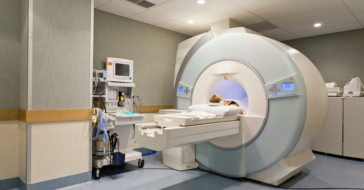
What Is a PSMA PET CT Scan?
A PSMA PET CT scan is a specialised imaging procedure used mainly for prostate cancer assessment. It combines two imaging technologies:
PET (Positron Emission Tomography)
Detects the distribution of a PSMA-targeting radioactive tracer within the body.
CT (Computed Tomography)
Provides anatomical images that help locate findings identified on PET imaging.
PSMA stands for Prostate-Specific Membrane Antigen, a protein found on the surface of many prostate cancer cells.
During the scan, a PSMA-targeting radiotracer is injected into a vein. The tracer circulates through the body and binds to tissues expressing PSMA. A PET CT scanner detects the tracer distribution and produces images that help identify suspicious lesions.
PSMA expression is not exclusive to prostate cancer. Some normal tissues, benign conditions and other tumours can also show PSMA tracer uptake.
The findings must therefore be interpreted by a qualified nuclear medicine physician alongside PSA levels, clinical history and other investigations.
Why Is a PSMA PET CT Scan Done?
A PSMA PET CT scan for prostate cancer provides information that may influence staging, treatment selection and follow-up.
1. Initial Prostate Cancer Staging
PSMA PET CT may be recommended for patients with newly diagnosed prostate cancer, particularly those with unfavourable intermediate-risk or high-risk disease.
It helps evaluate whether cancer has spread beyond the prostate to nearby lymph nodes, bones or other organs.
2. Detecting Prostate Cancer Recurrence
After prostate cancer treatment, a rising Prostate-Specific Antigen (PSA) level may indicate possible recurrence.
PSMA PET CT can help identify suspected recurrent disease after radical prostatectomy or radiation therapy, including lesions that may not be clearly visible on conventional imaging.
3. Evaluating Metastatic Prostate Cancer
PSMA PET CT helps assess the distribution of PSMA-expressing prostate cancer lesions in lymph nodes, bones and other tissues.
This information can assist specialists in determining the extent of disease and planning treatment.
4. Planning Prostate Cancer Treatment
The results may influence decisions regarding surgery, radiation therapy, systemic treatment or other management approaches.
Treatment decisions depend on the patient's overall clinical condition, cancer characteristics and multidisciplinary assessment.
5. Assessing Eligibility for PSMA-Targeted Therapy
In selected patients with advanced prostate cancer, PSMA PET imaging is used to evaluate PSMA expression before considering PSMA-targeted radioligand therapy.
Eligibility depends on the specific treatment, applicable clinical criteria and specialist evaluation.
When Is a PSMA PET CT Scan Recommended?
Doctors may recommend PSMA PET CT in the following clinical situations:
| Clinical situation | Purpose of the scan |
|---|---|
| Newly diagnosed higher-risk prostate cancer | Assess lymph node and distant metastatic disease |
| Rising PSA after prostate surgery | Investigate suspected biochemical recurrence |
| Rising PSA after radiation therapy | Evaluate possible recurrent disease |
| Suspected metastatic prostate cancer | Identify the location and extent of PSMA-expressing lesions |
| Planning selected salvage treatments | Help determine the distribution of recurrent disease |
| Evaluation before PSMA-targeted radioligand therapy | Assess tumour PSMA expression according to treatment-specific criteria |
Important: PSMA PET CT is not a routine screening test for prostate cancer and does not replace PSA assessment, prostate MRI or biopsy when these investigations are clinically required.
The referring specialist determines whether the scan is appropriate.
Types of PSMA PET CT Scans
Different PSMA-targeting radiotracers are used in clinical practice. The choice depends on availability, clinical indication and the imaging protocol.
1. Ga-68 PSMA PET CT Scan
Gallium-68-labelled PSMA tracers, including Ga-68 PSMA-11, are used to image PSMA-expressing tissues.
Ga-68 PSMA PET CT is commonly used for prostate cancer staging and suspected recurrence.
2. F-18 PSMA PET CT Scan
Fluorine-18-labelled PSMA tracers, such as F-18 DCFPyL and F-18 PSMA-1007, are also used in prostate cancer imaging.
Different F-18 tracers have distinct imaging characteristics and patterns of normal tissue uptake.
Both Ga-68 and F-18 PSMA tracers can provide clinically useful information. They are not identical, and the choice should follow the nuclear medicine team's protocol.
Note: Patients should confirm which PSMA radiotracer is available at the imaging facility before scheduling an appointment.
How Is a PSMA PET CT Scan Performed?
The procedure generally involves five stages.
Step 1: Registration and Medical Assessment
The imaging team reviews the patient's referral, prostate cancer diagnosis, PSA results, treatment history and previous imaging reports.
Any relevant medications, allergies or medical conditions should be discussed before the examination.
Step 2: PSMA Radiotracer Injection
A small amount of PSMA-targeting radioactive tracer is administered through an intravenous injection, usually in the arm.
The tracer circulates through the bloodstream and binds to tissues expressing PSMA.
Step 3: Tracer Uptake Period
After injection, the patient waits while the tracer distributes throughout the body.
For commonly used PSMA tracers, imaging often begins approximately 60 minutes after injection. The waiting period varies depending on the tracer and protocol.
Step 4: PET CT Imaging
The patient lies on a scanning table that moves through the PET CT scanner.
The scanner acquires PET and CT images, allowing the imaging team to assess tracer distribution and anatomical structures.
Patients are asked to remain still during scanning to reduce motion-related image artefacts.
Step 5: Image Interpretation
A qualified nuclear medicine physician reviews the images and prepares a report describing relevant PSMA uptake and CT findings.
The referring doctor interprets the results alongside PSA levels, biopsy findings and the patient's clinical condition.
How to Prepare for a PSMA PET CT Scan
Preparation for PSMA PET CT differs from FDG PET CT. In particular, routine fasting and blood glucose control are generally not required for standard PSMA imaging, unless the imaging centre specifies otherwise.
Before Your Scan
- Food and fasting: Fasting is usually not necessary for a standard PSMA PET CT scan. Follow the facility's instructions, especially if contrast-enhanced CT is planned.
- Hydration: Drink adequate water before and after the scan unless your doctor has restricted fluid intake.
- Medications: Continue regular medications unless your treating doctor or imaging team advises otherwise.
- Medical records: Carry recent PSA reports, biopsy or histopathology results, treatment summaries and previous imaging reports.
- Treatment history: Inform the imaging team about prostate surgery, radiation therapy, hormone therapy, chemotherapy or other prostate cancer treatments.
- Contrast precautions: Report any previous contrast reaction, kidney disease or other relevant medical conditions if CT contrast is planned.
- Clothing: Wear comfortable clothing and remove metallic objects as instructed.
Important: Patients should not independently stop hormone therapy or other cancer medications before the scan.
How Long Does a PSMA PET CT Scan Take?
A PSMA PET CT appointment commonly takes approximately 2–3 hours, although the total duration varies with the tracer, imaging protocol and individual requirements.
The appointment generally includes:
- Registration and clinical review
- Intravenous tracer injection
- Tracer uptake period, often approximately 60 minutes
- PET CT image acquisition, commonly around 20–40 minutes
Additional imaging or contrast-enhanced CT may increase the duration.
The imaging facility will provide the expected appointment time.
Is a PSMA PET CT Scan Safe?
PSMA PET CT is an established nuclear medicine imaging procedure that uses a controlled amount of radioactive tracer along with CT imaging.
The examination involves ionising radiation exposure from the PET tracer and the CT component.
The expected diagnostic benefit is considered against radiation exposure when deciding whether the examination is appropriate.
Important Safety Considerations
- Radiation exposure: The radiation dose depends on the administered tracer activity and CT protocol.
- Injection discomfort: Patients may experience brief discomfort when the intravenous injection is given.
- Contrast-related risks: If intravenous CT contrast is required, additional precautions may be necessary for patients with kidney problems or previous contrast reactions.
- Radiation precautions: The imaging team may advise temporary precautions following tracer administration, depending on the protocol.
- Repeat examinations: The need and timing of repeat PSMA PET CT scans should be determined by the treating specialist.
PSMA PET CT should be performed only when medically indicated and under appropriate nuclear medicine supervision.
How Accurate Is a PSMA PET CT Scan?
PSMA PET CT can detect prostate cancer lesions that may not be visible on conventional imaging, particularly in selected patients with suspected recurrence.
However, its accuracy varies according to several factors:
- PSA level at the time of scanning
- Size and location of lesions
- PSMA expression in the tumour
- Type of radiotracer used
- Previous cancer treatments
- Timing and quality of imaging
Small lesions may remain undetected, and some prostate cancers show low or absent PSMA expression.
Additionally, PSMA uptake can occur in certain benign conditions and non-prostate tumours.
A negative PSMA PET CT scan does not completely exclude prostate cancer or recurrence. Similarly, a positive finding may require correlation with other imaging or tissue sampling.
PSMA PET CT Scan vs FDG PET CT Scan
Although both examinations use PET CT technology, they assess different biological characteristics.
| Feature | PSMA PET CT Scan | FDG PET CT Scan |
|---|---|---|
| Main target | PSMA expression | Glucose metabolism |
| Common tracers | Ga-68 or F-18 PSMA tracers | F-18 FDG |
| Primary cancer application | Prostate cancer | Several cancer types |
| Typical clinical uses | Prostate cancer staging, recurrence and selected treatment planning | Cancer staging, treatment response and recurrence assessment |
| Routine fasting | Usually not required | Generally required |
| Blood glucose assessment | Not routinely necessary | Commonly required |
| Main limitation | Tumours with low PSMA expression may be missed | Tumours with low FDG uptake may be missed |
PSMA PET CT is generally preferred over FDG PET CT for many established prostate cancer imaging indications. However, FDG PET CT may provide additional information in selected aggressive or advanced prostate cancers.
The appropriate examination depends on the clinical indication.
What Happens After a PSMA PET CT Scan?
Most patients can return to normal daily activities after the examination unless they have received sedation or specific instructions.
Patients are generally encouraged to drink water and empty their bladder regularly to help eliminate the radiotracer.
The imaging team may provide temporary radiation safety instructions based on the tracer used.
The scan report describes areas of PSMA uptake and their anatomical locations.
The treating urologist or oncologist reviews the results alongside PSA levels, biopsy findings, previous treatments and other investigations to determine the next steps.
An abnormal PSMA PET CT finding does not automatically establish the final diagnosis or treatment plan.
PSMA PET CT Scan in Navi Mumbai – Appointment Information
Patients referred for a PSMA PET CT Scan in Navi Mumbai should confirm the imaging requirements and tracer availability before scheduling an appointment.
It is useful to keep the referring doctor's prescription, recent PSA results, prostate biopsy reports and previous imaging records ready.
Patients should also inform the imaging team about previous prostate cancer treatments, including surgery, radiation therapy and hormonal treatment.
For PSMA PET CT scan-related enquiries in Navi Mumbai, contact the diagnostic service to confirm tracer availability, appointment requirements, preparation instructions and reporting details.
The final imaging protocol is determined by the nuclear medicine team based on the patient's clinical requirements.
Frequently Asked Questions About PSMA PET CT Scan
PSMA PET CT is primarily used for prostate cancer staging, evaluation of suspected recurrence and assessment of metastatic disease. It also helps determine eligibility for certain PSMA-targeted treatments.
PSMA PET CT can identify many prostate cancer lesions that express PSMA. However, it cannot detect every tumour and does not replace prostate biopsy for establishing an initial diagnosis.
Fasting is generally not required for standard PSMA PET CT imaging. However, specific preparation may be necessary if contrast-enhanced CT or another procedure is planned.
The complete appointment commonly takes approximately 2–3 hours, including tracer injection, uptake time and image acquisition.
The imaging procedure itself is generally painless. There may be brief discomfort from the intravenous injection.
Both use radiotracers that target PSMA. They differ in radioactive isotope, tracer characteristics and imaging protocols. The appropriate tracer depends on clinical requirements and availability.
Yes. PSMA PET CT can identify many PSMA-expressing prostate cancer deposits in bones, lymph nodes and other organs. However, a negative scan does not exclude all metastatic disease.
PSMA PET CT can detect recurrent prostate cancer in some patients with low PSA levels. Detection rates generally increase as PSA levels rise, although results vary between patients.
PSMA PET CT is generally more appropriate for many prostate cancer staging and recurrence indications. FDG PET CT may be useful in selected aggressive or advanced cases.
PSMA PET CT is generally well tolerated when performed for a suitable medical indication. It involves ionising radiation, and the medical team considers the expected diagnostic benefit and potential risks.
No. Small lesions and tumours with low PSMA expression may not be detected. The scan findings must be interpreted alongside PSA results and other investigations.
PSMA PET CT should be performed for an appropriate clinical indication following medical evaluation. Patients should provide a referral or clinical recommendation as required by the imaging facility.
Enquire About a PSMA PET CT Scan in Navi Mumbai
If your treating doctor has recommended a PSMA PET CT scan, confirm the radiotracer, preparation instructions, appointment availability and required documents before visiting the imaging facility.
Contact the diagnostic service for PSMA PET CT scan information and appointment enquiries.
Medical disclaimer: This content is intended for general patient education and does not replace consultation with a qualified urologist, oncologist or nuclear medicine physician. The suitability of PSMA PET CT depends on individual clinical circumstances.