
Being diagnosed with prostate cancer can bring several questions. Has the cancer remained within the prostate? Has it reached nearby lymph nodes or bones? If treatment has already been completed, why is the PSA level rising again?
A PSMA PET CT scan for prostate cancer can help doctors answer some of these questions by identifying areas that may contain prostate cancer cells.
Unlike a routine CT scan, PSMA PET-CT uses a specialised radioactive tracer that targets a protein commonly found in higher amounts on prostate cancer cells.
However, not everyone diagnosed with prostate cancer needs this examination. The decision depends on the cancer's risk category, PSA levels, previous treatment and whether the scan findings are likely to change the treatment plan.
This guide explains when a PSMA PET-CT scan may be recommended, how the examination works, what the results mean and what patients should know before scheduling a scan in Navi Mumbai.
What Is a PSMA PET CT Scan?
Quick Answer: A PSMA PET-CT scan is a specialised nuclear medicine examination used to locate prostate cancer in selected patients. It combines molecular imaging with CT pictures to help assess whether cancer has spread or returned after treatment.
PSMA stands for Prostate-Specific Membrane Antigen, a protein expressed by many prostate cancer cells.
During the examination, a small amount of a radioactive substance, called a radiotracer, is injected into a vein. The tracer binds to PSMA-expressing tissue, allowing the PET scanner to detect areas of tracer accumulation.
The CT component provides anatomical information, helping doctors identify where these areas are located.
Common PSMA radiotracers include:
- Gallium-68 (Ga-68) PSMA: A radiotracer used for prostate cancer imaging.
- Fluorine-18 (F-18) PSMA: Another category of PSMA-targeted radiotracers used in specialised PET imaging.
Both are designed to help visualise PSMA-expressing tissue, although their imaging characteristics and availability differ.
Important: PSMA uptake is not exclusive to prostate cancer. Some normal tissues, benign conditions and other diseases can also show tracer uptake. Results must be interpreted alongside the patient's clinical history and other investigations.
When Is a PSMA PET CT Scan Recommended for Prostate Cancer?
A PSMA PET-CT scan is particularly useful when information about the location or spread of prostate cancer could influence treatment decisions.
1. Initial Staging of High-Risk Prostate Cancer
After a prostate cancer diagnosis, doctors assess the risk of the disease spreading beyond the prostate.
This evaluation usually includes PSA levels, biopsy findings, Gleason score or ISUP Grade Group, and the clinical stage.
For patients diagnosed with high-risk or certain intermediate-risk prostate cancers, a PSMA PET-CT scan may be recommended to look for cancer outside the prostate.
The examination may identify:
- Cancer involving nearby pelvic lymph nodes.
- Cancer spread to bones.
- Disease in distant lymph nodes.
- Other sites of suspected metastatic disease.
These findings can influence decisions about surgery, radiation therapy and systemic treatment.
The 2026 European Association of Urology (EAU) guidelines support PSMA PET-CT for high-risk staging and recommend considering it for unfavourable intermediate-risk disease, if available.
2. Rising PSA After Prostate Cancer Surgery
After radical prostatectomy, the prostate gland is removed. PSA is expected to fall to very low or undetectable levels.
If PSA remains detectable or begins rising after surgery, the doctor may investigate possible persistent or recurrent disease.
This is one of the important situations in which a PSMA PET scan after prostate surgery may be helpful.
The scan can assist in evaluating whether cancer is present in the prostate bed, nearby lymph nodes or more distant areas.
For example, a patient whose PSA begins rising after surgery may undergo PSMA PET-CT to help determine whether local salvage radiation or another treatment approach should be considered.
Current European guidance recommends considering PSMA PET-CT after prostatectomy when PSA exceeds 0.2 ng/mL and the results are expected to influence treatment decisions. However, scan timing must be individualised, and a negative scan should not automatically delay otherwise appropriate salvage treatment.
3. Suspected Recurrence After Radiation Therapy
PSA behaves differently after radiation therapy because the prostate gland usually remains in place.
PSA may decrease gradually rather than becoming undetectable.
If PSA rises in a pattern suggesting recurrence, doctors may recommend further evaluation.
A PSMA PET-CT scan may help identify whether the disease has returned within the prostate, affected nearby lymph nodes or spread elsewhere.
The results may guide discussion about additional local treatment or systemic therapies.
Not every temporary PSA increase means cancer has returned. PSA trends, previous treatment and clinical findings remain important.
4. When CT or Bone Scans Are Negative or Unclear
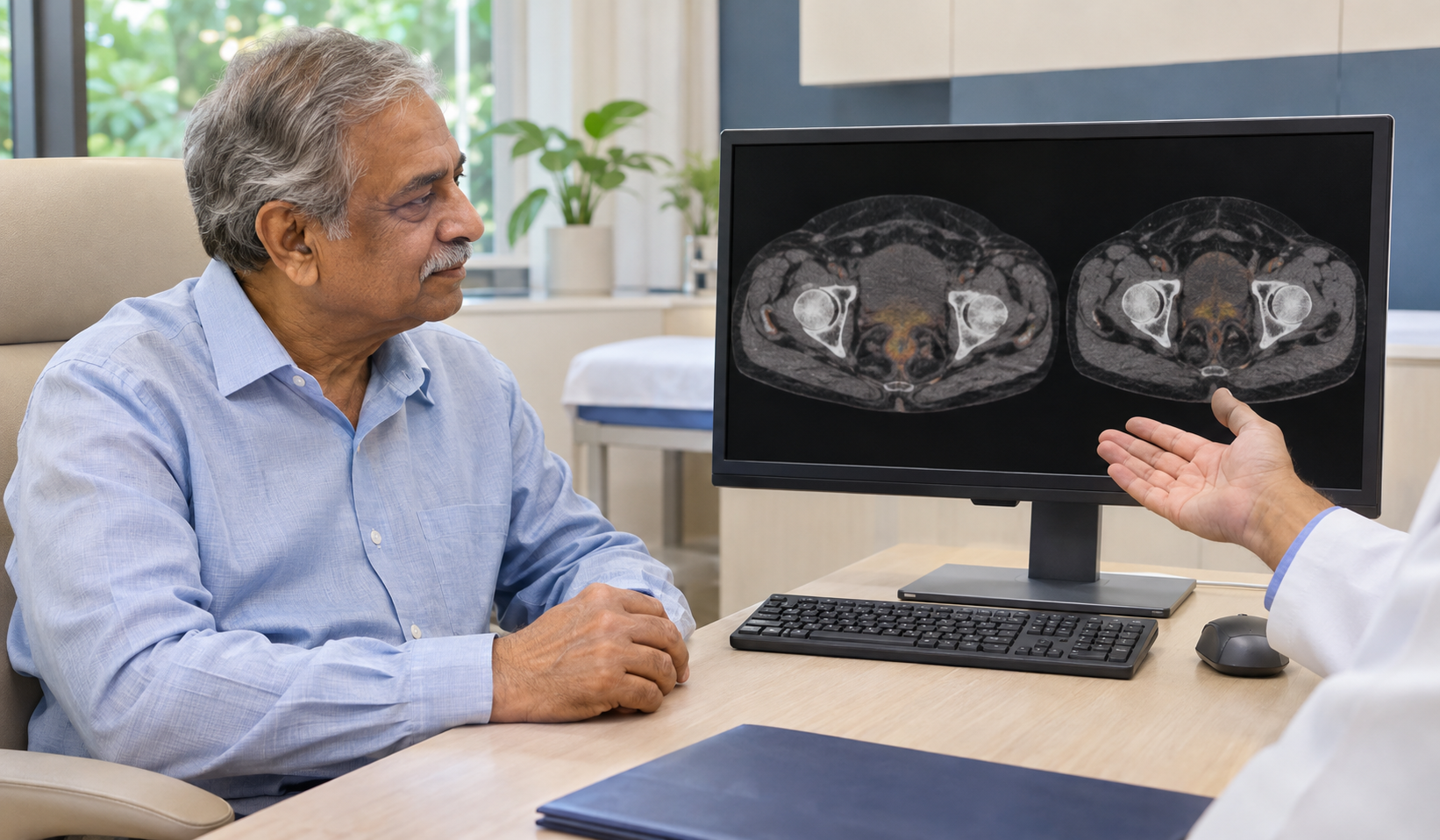
Conventional imaging, such as CT and bone scans, may not detect all small areas of prostate cancer.
In selected patients, PSMA PET-CT may identify suspected disease that is not visible on conventional investigations.
For example, a patient may have a rising PSA level while their CT and bone scan reports show no definite evidence of cancer spread.
A PSMA PET-CT scan may provide additional information that helps the treating doctor plan further management.
Studies have demonstrated improved staging accuracy with PSMA PET-CT compared with conventional CT and bone scans in high-risk prostate cancer. Nevertheless, very small cancer deposits may remain undetected.
5. Evaluating Advanced or Metastatic Prostate Cancer
Patients with advanced prostate cancer may require imaging to establish the location and extent of disease.
A PSMA PET-CT scan may be considered in specific circumstances to clarify disease distribution or support treatment decisions.
Doctors may use the findings alongside PSA trends, symptoms, CT, MRI and other tests.
It is not necessarily the appropriate routine monitoring scan for every patient receiving treatment.
6. Before Certain PSMA-Targeted Treatments
In selected patients with advanced prostate cancer, PSMA PET imaging may help assess whether tumours express enough PSMA to meet the imaging criteria for PSMA-targeted radioligand therapy.
One example is lutetium-177 PSMA treatment, used in appropriately selected patients.
A positive scan alone does not establish eligibility. Doctors must also consider prior treatments, the disease setting, overall health and relevant treatment criteria.
Is a PSMA PET CT Scan Necessary for Every Prostate Cancer Patient?
No. A PSMA PET-CT scan is not routinely necessary for every patient diagnosed with prostate cancer.
Patients with low-risk prostate cancer who have no clinical indication of spread usually do not require PSMA PET-CT for initial staging.
For some patients, PSA testing, prostate MRI, biopsy findings and clinical assessment may provide sufficient information for initial management.
| Clinical situation | Is PSMA PET-CT commonly considered? |
|---|---|
| Elevated PSA without a confirmed cancer diagnosis | Generally not as an initial diagnostic test |
| Confirmed low-risk prostate cancer | Usually not routinely indicated |
| Unfavourable intermediate-risk prostate cancer | May be recommended |
| High-risk prostate cancer | Commonly recommended for staging |
| Rising PSA after radical prostatectomy | Often considered if results will guide treatment |
| Suspected recurrence after radiation | Often considered |
| Advanced cancer requiring treatment selection | May be appropriate in selected cases |
These are general clinical situations rather than fixed rules. The treating specialist determines whether the examination is appropriate.
How Does PSMA PET CT Help Detect Prostate Cancer Spread?
Prostate cancer may spread through lymphatic channels or the bloodstream.
Common sites of metastasis include pelvic lymph nodes and bones.
PSMA PET-CT detects areas of PSMA-targeted tracer uptake, while CT helps determine their anatomical location.
This can assist in identifying suspected disease in areas such as the pelvis, spine, ribs or distant lymph nodes.
However, a PSMA PET-CT scan cannot guarantee detection of every cancer cell. Microscopic deposits may be too small to identify, and some prostate cancers show relatively low PSMA expression.
A suspicious scan finding may require correlation with MRI, follow-up imaging or occasionally a biopsy.
PSMA PET CT vs FDG PET CT: What Is the Difference?
PSMA PET-CT and FDG PET-CT are nuclear medicine imaging examinations, but they use different radiotracers and provide different biological information.
| Feature | PSMA PET-CT | FDG PET-CT |
|---|---|---|
| Main target | PSMA protein expression | Glucose metabolism |
| Radiotracer | Ga-68 or F-18 PSMA tracer | F-18 FDG |
| Prostate cancer use | Commonly used for staging and recurrence evaluation | Selected cases, particularly certain aggressive cancers |
| Primary purpose | Identify PSMA-expressing disease | Identify metabolically active tissue |
| Recommended for every patient? | No | No |
PSMA PET-CT is often more suitable than FDG PET-CT for evaluating typical prostate adenocarcinoma.
However, FDG PET-CT may have a role in selected aggressive or advanced cancers, particularly when tumour biology suggests reduced PSMA expression.
The treating specialist determines which examination is appropriate.
What Happens During a PSMA PET CT Scan?
The examination is generally performed as an outpatient procedure and does not involve surgery.
-
Step 1: Registration and Clinical Review
The imaging team reviews the prescription, relevant medical history, PSA reports, previous scans and prostate cancer treatment details.
-
Step 2: Radiotracer Injection
A small amount of PSMA-targeted radioactive tracer is injected into a vein, usually in the arm.
The injection feels similar to a routine blood test.
-
Step 3: Tracer Uptake Period
After the injection, patients wait while the tracer circulates and accumulates in relevant tissues.
For many commonly used PSMA radiotracers, this waiting period is approximately 60 minutes. Some tracers require a longer interval.
-
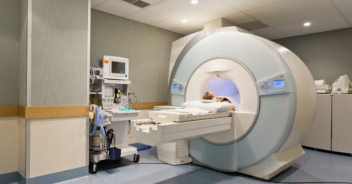
Step 4: PET CT Imaging
The patient lies on a scanning table that moves through the scanner.
Remaining still helps produce clear images. The scan itself is painless.
-
Step 5: Image Interpretation
The PET and CT images are processed and reviewed by a qualified nuclear medicine specialist.
The report is then provided to the referring doctor to guide further evaluation or treatment planning.
How long does a PSMA PET CT scan take?
Patients may need approximately 2–3 hours for the complete appointment, although this varies with the tracer, imaging protocol and whether additional images are required.
How Should You Prepare for a PSMA PET CT Scan?
Preparation depends on the radiotracer and the examination protocol.
Unlike a standard FDG PET-CT scan, fasting is not routinely required for many PSMA PET-CT protocols. However, patients should follow the specific instructions provided by their imaging centre. You can also read our detailed guide on PET CT scan preparation.
Before the appointment:
- Bring your doctor's prescription and recent PSA reports.
- Carry previous PET-CT, CT or MRI scans, if available.
- Bring your prostate biopsy or histopathology report.
- Inform the team about previous surgery, radiation or hormonal treatment.
- Mention kidney problems, allergies and any recent treatment changes.
- Follow the centre's instructions about drinking water and emptying your bladder.
If contrast-enhanced CT is planned, additional preparation may be necessary.
Do not stop prescribed medicines or hormonal treatment unless your treating doctor specifically advises it.
What Do PSMA PET CT Scan Results Mean?
A PSMA PET-CT report describes the location and intensity of radiotracer uptake and whether the findings are suspicious for prostate cancer.
The findings may include:
No suspicious PSMA uptake
No definite PSMA-avid cancer is identified. However, microscopic disease or cancer with low PSMA expression cannot be excluded.
Uptake confined to the prostate or prostate bed
This may indicate disease within the prostate or possible local recurrence after treatment, depending on the patient's history.
Uptake in pelvic lymph nodes
This may suggest regional lymph node involvement.
Uptake in bones or distant organs
These findings may raise concern about metastatic disease and require interpretation in the clinical context.
A report may also use the term SUV (Standardised Uptake Value), which describes the measured concentration of tracer activity in a particular area.
A higher SUV does not automatically establish that an abnormality is cancerous or indicate how aggressive it is.
The scan findings should be assessed alongside PSA trends, biopsy results, previous imaging and clinical examination.
Can a PSMA PET CT Scan Detect Prostate Cancer at Low PSA Levels?
Yes. PSMA PET-CT can sometimes identify recurrent prostate cancer when PSA levels are relatively low, including cases where conventional imaging is negative.
However, the probability of detecting disease generally increases as PSA levels rise.
Detection also depends on factors such as PSA doubling time, tumour characteristics, tracer type and lesion size.
A negative PSMA PET-CT scan at a low PSA level does not necessarily mean that recurrent cancer is absent.
For patients being evaluated for salvage radiation therapy after prostatectomy, treatment decisions should not be based on the scan result alone.
Is a PSMA PET CT Scan Safe?
PSMA PET-CT uses a small amount of radioactive tracer and involves exposure to ionising radiation from both PET and CT components.
The examination is generally well tolerated when appropriately prescribed and performed.
Possible considerations include minor injection-site discomfort, uncommon tracer-related reactions and any additional risks associated with intravenous CT contrast, if used.
The imaging team follows radiation safety procedures and provides relevant precautions after the scan.
Because the examination involves radiation exposure, it should be performed when the expected clinical benefit justifies the investigation.

PSMA PET CT Scan Cost in Navi Mumbai
The PSMA PET CT scan cost in Navi Mumbai depends on the selected radiotracer, imaging protocol, diagnostic centre and additional services.
Factors that may influence the charges include the use of Gallium-68 or Fluorine-18 radiotracers, scan coverage, contrast-enhanced CT requirements and reporting arrangements.
Before booking, patients should confirm whether the quotation includes the radiotracer, examination, specialist report and digital images.
For more information about pricing factors, read our guide on PET CT scanning in Navi Mumbai and enquire about the prescribed PSMA examination.
PSMA PET CT Scan in Navi Mumbai: What Should Patients Consider?
Patients referred for prostate cancer imaging in Navi Mumbai may look for PSMA PET-CT services in CBD Belapur, Vashi, Nerul and surrounding areas.
Before scheduling the examination, confirm that the imaging centre provides the exact tracer and protocol requested by the treating doctor.
It is also useful to check preparation instructions, report availability, the required medical documents and total charges.
For information about a PSMA PET CT scan in Navi Mumbai, patients can contact PET Scan in Navi Mumbai, CBD Belapur, with their doctor's prescription to enquire about the relevant examination and current availability.
Frequently Asked Questions About PSMA PET CT Scan
1. Why is a PSMA PET CT scan recommended for prostate cancer?
A PSMA PET-CT scan may be recommended to assess cancer spread at diagnosis, investigate suspected recurrence after treatment or guide selected treatment decisions.
2. Is PSMA PET CT necessary for every prostate cancer patient?
No. It is generally more useful in high-risk prostate cancer, selected intermediate-risk cases and suspected recurrence. Routine staging is usually unnecessary for low-risk disease.
3. Can PSMA PET CT detect prostate cancer after surgery?
Yes. It may help locate suspected recurrent or persistent prostate cancer when PSA remains detectable or rises after radical prostatectomy.
4. At what PSA level is a PSMA PET CT scan recommended?
There is no universal PSA threshold for every patient. After prostatectomy, European guidance recommends considering imaging when PSA is above 0.2 ng/mL if the findings will influence treatment decisions. Individual circumstances may justify a different approach.
5. Can PSMA PET CT detect prostate cancer in bones?
Yes. PSMA PET-CT can identify suspicious PSMA-expressing lesions in bones, although not every bone abnormality represents metastatic cancer.
6. Does a negative PSMA PET CT mean there is no prostate cancer?
No. A negative scan does not completely exclude microscopic cancer or disease with limited PSMA expression.
7. Is PSMA PET CT painful?
The scan itself is painless. Patients may experience mild discomfort from the intravenous tracer injection.
8. Is fasting required before PSMA PET CT?
Fasting is not routinely necessary for many PSMA PET-CT protocols. Follow the instructions provided by the imaging centre, especially if additional investigations are planned.
9. What is the difference between Ga-68 PSMA and F-18 PSMA scans?
Both use PSMA-targeted radiotracers. They differ in radioactive isotope, production requirements and imaging characteristics. The choice depends on the clinical indication and available protocol.
10. How much does a PSMA PET CT scan cost in Navi Mumbai?
Charges vary according to the tracer, diagnostic centre and examination requirements. Patients should request a current quotation before scheduling the scan.
11. Can PSMA PET CT replace a prostate biopsy?
No. PSMA PET-CT does not routinely replace prostate biopsy for establishing the initial diagnosis of prostate cancer. Biopsy provides essential tissue information for diagnosis and grading.
12. How soon will the PSMA PET CT report be available?
Reporting times vary by centre and examination complexity. Patients should confirm the expected reporting schedule when booking.
Conclusion
A PSMA PET CT scan for prostate cancer is an important imaging examination that can help doctors evaluate disease spread, investigate rising PSA levels after treatment and make informed management decisions.
It is particularly relevant for selected patients with high-risk prostate cancer or suspected recurrence, but it is not routinely necessary for everyone.
The decision to undergo the examination should be based on the patient's PSA levels, biopsy results, previous treatment and overall clinical assessment.
Planning a PSMA PET CT scan in Navi Mumbai? Contact PET Scan in Navi Mumbai, CBD Belapur, with your doctor's prescription to enquire about tracer availability, preparation instructions, appointment scheduling and applicable charges.
You can also learn more about our centre or read patient testimonials.
Planning a PSMA PET CT scan in Navi Mumbai?
Contact the imaging centre with your doctor's prescription to enquire about tracer availability, preparation instructions, appointment scheduling and applicable charges.
Medical Disclaimer: This article is intended for general educational purposes and does not replace individual medical advice. PSMA PET-CT should be performed when clinically indicated and recommended by an appropriately qualified medical professional.